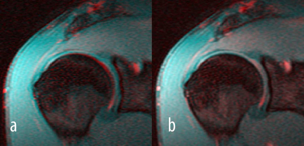
Figure 5.
Shown are colour-coded images of the fourth (red) and first (blue) echo of a T2 acquisition before registration a) and after registration b). In particular around the subchondral bone a clear misalignment can be seen between the two echoes in a) which disappeared after aligning the fourth to the first echo in b).

