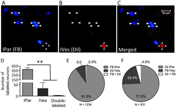
Figure 1. IPar- and IVes-labeled neurons are anatomically distinct subsets of PN bladder afferents.
Intraparynchemal (IPar) injection of FB and intravesical (IVes) infusion of DiI retrogradely labeled two subpopulations of bladder-innervating L6 DRG neurons (A-C). Arrowheads indicate IPar neurons labeled by FB and asterisks indicate a DiI-positive, IVes neuron. One wk after dye administration, the total number of L6 DRG neurons labeled by IPar FB was significantly higher than the number labeled by IVes DiI or by both FB and DiI (D). Of a total of 1234 labeled L6 DRG neurons, 91.5% were positive for FB, 8% were positive for DiI, and 0.5% were double-labeled (E). When dyes were reversed, 77.6% of IPar-labeled neurons were positive for DiI, 22.4% IVes-labeled neurons were positive for FB, and 4.5% were double-labeled out of a total of 531 labeled neurons (F). ** indicates P<0.01.

