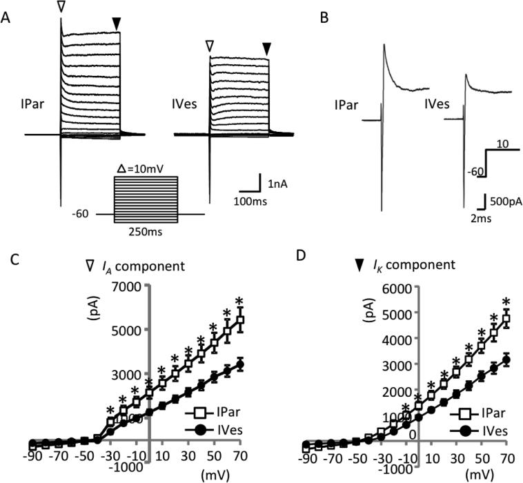
Figure 5. Magnitude of outward currents differs between urothelial and non-urothelial PN bladder neurons.
Representative traces of total currents evoked by voltage steps (shown below the traces) from a holding potential of −73 mV revealed an inactivating inward current, an inactivating outward current (white triangles), and a non-inactivating outward current (black triangles) (A). The inactivating outward currents measured in non-urothelial (IPar-labeled) neurons were significantly higher at voltage steps ranging from −43 to +57 mV than in urothelial (IVes-labeled) neurons (B,C). Similarly, the non-inactivating outward current measured in non-urothelial relative to urothelial neurons was significantly higher at voltage steps of −23 to +57 mV (D). N=10 neurons obtained from 3 animals/group. * indicates P<0.05.

