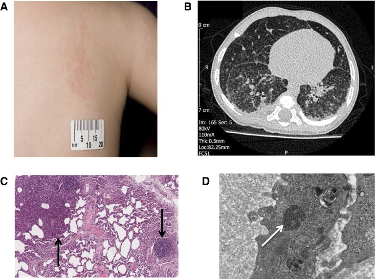
Figure 1.
(A) Raised, papular, erythematous rash on the trunk. (B) Chest computed tomography scan showing widespread, slightly nodular interstitial opacification in both lungs. There are a number of small (<1 cm) peripheral cysts within the right lower and middle lobes. (C) Light microscopy microphotograph of lung biopsy (low magnification, hematoxylin and eosin stain), showing solid area composed of mixed infiltrate (left arrow) and chronic inflammation (right arrow). (D) Electron microscopy photograph of lung endothelium, showing tuboreticular inclusions (arrow).

