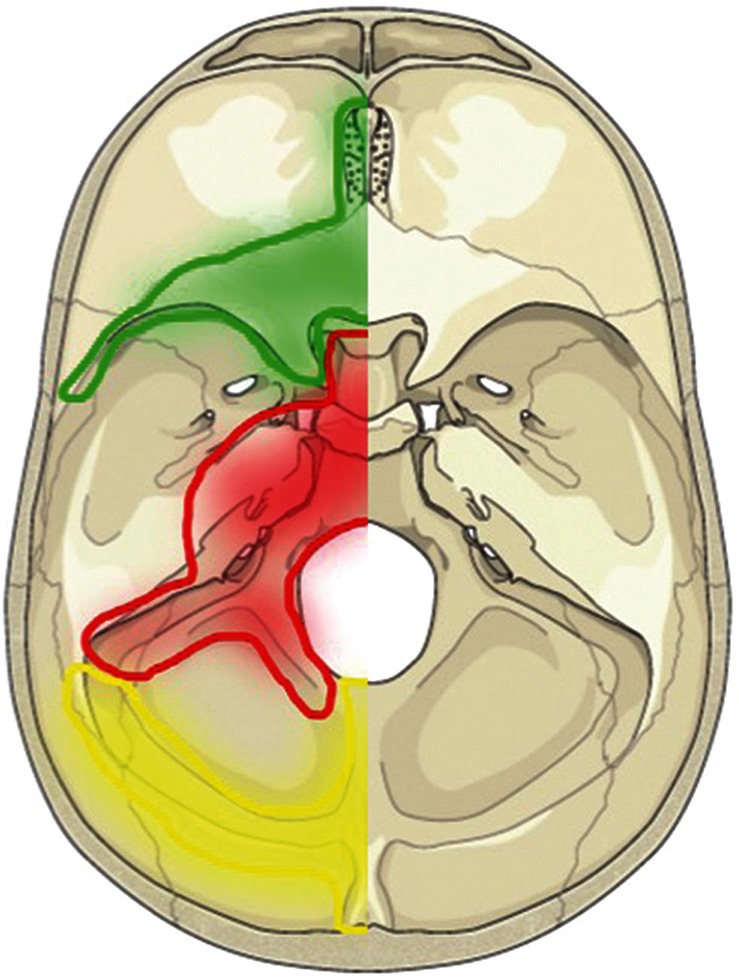
Fig. 1 .

Topographical map of the three different groups of DAVFs in relation to the embryological domains and bony structures. The green area depicts the falx and the tent of the cerebellum (FT group) derived from neural crest cells. The red area reveals the ventral group on the surface of endochondral bone (VE group). The yellow area depicts the torcular, transverse sinus, medial occipital sinus, and posterior marginal sinus that are associated with the surface of membranous bone (DM group).
