Figure 2.
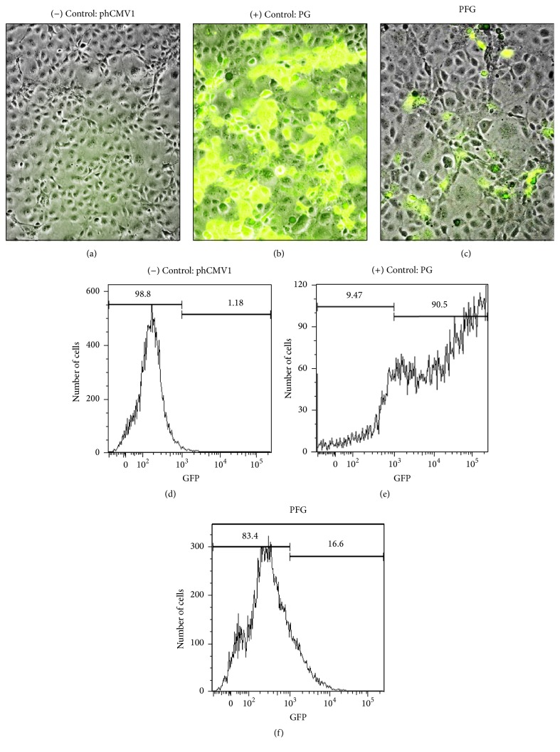
Expression of RSV F protein in Cos-7 cells. Visual and quantitative analyses demonstrated that RSV F protein was expressed in vitro. Immunofluorescence microscopy of (a) phCMV1 (negative control), (b) phCMV1+GFP (positive control), and (c) PFG (DNA vaccine) transfected cells. Flow cytometric analysis of transfected cells: (d) Cos-7 cells (negative control), (e) phCMV1+GFP (positive control), and (f) PFG (DNA vaccine).

