
Figure 3.
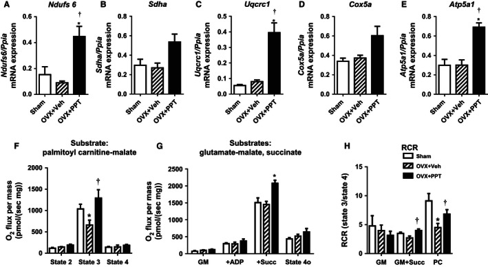
PPT treatment enhances cardiac mitochondrial function. Cardiac mRNA expression of the (A–E) mitochondrial genes Ndufs6, Sdha, Uqcrc1, Cox5a, Atp5a1 were measured by quantitative RT‐PCR. (Data shown as mean ± SEM; N = 3 sham, N = 4 OVX + Veh, and N = 6 OVX + PPT. *P < 0.05 vs. sham, †P < 0.05 vs. OVX + Veh by Mann–Whitney test). Oxygen consumption rates of cardiac mitochondrial isolated from sham, OVX + Veh, and OVX + PPT‐treated mice. (F) Respiration rates for palmitoyl carnitine‐malate (PC) were measured at state 2, state 3, and state 4; (G) Respiration of cardiac with glutamate‐malate alone (GM; state 2 ADP‐independent), state 3 ADP‐supported respiration with GM + ADP (+ADP) and succinate (+Succ), and state 4o ATPase‐independent respiration after oligomycin addition; (H) Respiratory control ratios (RCR) were calculated for GM, GM + Succ, and PC. (Data shown as mean ± SEM; N = 4 for sham, OVX + Veh, and OVX + PPT. *P < 0.05 vs. sham, †P < 0.05 vs. OVX + Veh by Mann–Whitney test).