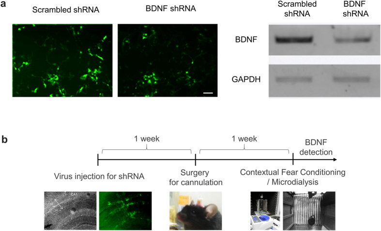
Figure 3. Development of anti-BDNF shRNA and in vivo gene silencing of BDNF using lentivirus.
(a) Validation of BDNF shRNA. Upper panel shows exemplary images of cultured HEK293T cells transfected with SC or BDNF shRNA candidates. mCherry and GFP were used as fluorescent reporters for BDNF and shRNA expression, respectively. Scale bar (white dash) indicates 30 μm. Middle panel shows result of RT-PCR of BDNF mRNA for each shRNA candidate. Lower panel shows bar graph of knockdown efficiency for each BDNF shRNA candidate. (b) Experimental procedure for BDNF detection from an animal in vivo.

