Fig. 2.
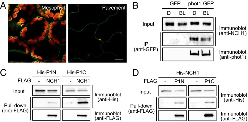
(A) Plasma-membrane localization of NCH1–GFP. Images are false colored to show GFP (green) and chlorophyll (red) fluorescence. (Scale bars, 10 µm.) (B) Coimmunoprecipitation of NCH1 with phot1. Microsomal fraction of the proteins from 10-d-old light-grown seedlings of transgenic GFP and phot1–GFP lines probed with anti-NCH1 or anti-phot1 antibodies. (C and D) Pull-down assay of phot1 and NCH1. FLAG- or His-tagged phot1 or NCH1 proteins were synthesized by in vitro transcription/translation reactions. The proteins bound to anti-FLAG beads were detected using anti-FLAG and anti-His.

