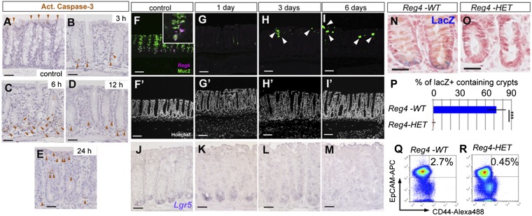
Fig. 3.
Lgr5+ colonic stem cells are lost upon ablation of DCS cells. (A–E) Active caspase-3 staining on DT-injected Reg4+/+ control mouse (A, n = 8 mice) and DT-injected Reg4DTR-Red mice at 3 h (B, n = 2 mice), 6 h (C, n = 2 mice), 12 h (D, n = 2 mice), and 24 h (E, n = 2). Apoptic cells were detected at the bottoms of crypts after administration of DT in Reg4DTR-Red mice (brown arrowheads in B–E), but not seen in control mice (A). (F–I) Daily DT administration for up to 6 d deleted DCS cells completely from colonic crypts. (F) Reg4 (magenta) and Muc2 (green) double positive-DCS cells were detected at the bottom part of crypt in DT-treated control Reg4+/+ mouse for 6 d (n = 6 mice). (G) One shot of DT eliminated all DCS cells and Goblet cells within 24 h (n = 6 mice). (H and I) DT treatment for 3 d (H, n = 4 mice) and 6 d (I, n = 4 mice) prevented reemergence of Reg4+ DCS cells, but a few goblet cells reappeared (white arrowheads). (F′–I′) Nuclei are stained with Hoechst (white). (J–M) Representative Lgr5 mRNA expression detected by a classical in situ hybridization in Reg4DTR-Red/+ mice. Lgr5 was present at the crypt bottoms of control (J, n = 8 mice) and after 1 d of DT treatment (K, n = 4 mice). The Lgr5 expression level became weaker after 3 d of DT (L, n = 4 mice) and was hardly detected for 6 d of DT treatment (M, n = 4 mice). (N–P) Visualization of stem cells in Lgr5-lacZ reporter mice in colonic crypts. Expression of lacZ was located at crypt base in Reg4+/+::Lgr5-lacZ (Reg4-WT) control mice 6 d after DT administration (N, n = 3 mice). No lacZ-positive cells were observed in Reg4DTR-Red/+::Lgr5-lacZ (Reg4-HET) mice 6 d after DT injection (O, n = 3 mice). (P) Quantification of Lgr5-lacZ–positive cells containing crypts in Reg4+/+::Lgr5-lacZ (Reg4-WT) control (n = 694 independent crypts in 4 mice) and Reg4DTR-Red/+::Lgr5-lacZ (Reg4-HET) (n = 1,087 independent crypts in 4 mice). (Q and R) FACS analysis of CD44+/EpCAM+ stem cell population from colonic crypts of Reg4-WT (Q, n = 3) control and Reg4-HET mice (R, n = 3) 6 d after DT injection. (Scale bars: A–E and J–M, 50 µm; F–I, 100 µm.)

