Fig. 6.
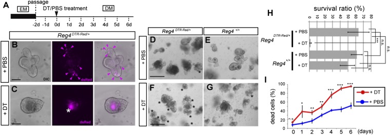
DCS cells are required for maintenance of colon organoids. (A) Dosing regimen used to study the elimination of Reg4+ cells in Reg4DTR-Red/+ colonic organoid. DM, differentiation medium; EM, expansion medium. (B and C) Organoids derived from isolated colonic crypts of Reg4DTR-Red/+ mouse were cultured in differentiation medium with PBS (B) or 67 ng⋅mL−1 DT (C) for 3 d. The expression of dsRed (magenta) was observed at budding structures in PBS-treated organoids (B, arrowheads) and inside lumen in DT-treated organoids (as auto-fluorescent background signal of apoptotic cells, asterisk) (C). Images are representative of four independent experiments. (D–G) Effect of deletion of Reg4+ DCS cell in colonic organoid derived from Reg4DTR-Red/+ (D and F) and wild type (E and G). Organoids after treatment with DT (F and G) or PBS (D and E) for 6 d. Asterisks indicate dead organoids. Reg4DTR-Red/+ treated with DT organoids were lost. Images are representative of four wells as biological replicates and repeated in two independent experiments. (H) The survival ratios of each organoids are shown (mean ± SD for four wells as biological replicates and repeated in two independent experiments) by two-way analysis of variance (ANOVA). (I) The percentage of dead cells of Reg4DTR-Red/+ organoids counted by FACS using propidium iodide in the presence of DT (red) and PBS (blue). Data are representative of four times technical repeated with mean ± SD (two-tailed Student's t test). *P < 0.05, **P < 0.01, ***P < 0.001, n.s., not significant. (Scale bars: B and C, 20 µm; D–G, 100 µm.)

