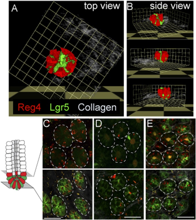
Fig. S3.
Relationship between Reg4+ DCS cells and Lgr5+ stem cells in each part of colon. (A and B) Three-dimension reconstruct of Lgr5+ GFP (green) and Reg4+ dsRed (red) in crypt bottom of middle-colon from lateral projection of a z stack and representative views from top (A) and side (B). (C–E) Intravital imaging of Reg4+ DCS (red) and Lgr5+ GFP (green) in a different region of colon, cecum (C), proximal (D), and distal (E), respectively. (Scale bars: C–E, 50 µm.)

