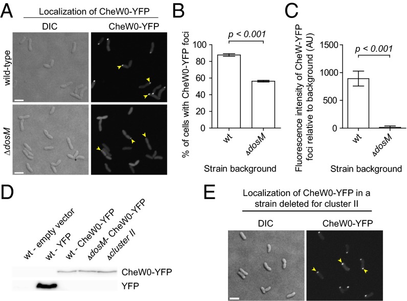
Fig. 3.
MCP DosM is required for the formation and stability of cluster I chemotactic signaling arrays. (A) Fluorescence micrograph showing the intracellular localization of CheW0-YFP in V. cholerae WT and ΔdosM strains. Yellow arrowheads indicate polar CheW0-YFP foci. (B) Bar graphs depicting the percentage of V. cholerae WT and ΔdosM cells with foci of CheW0-YFP. (C) Bar graphs representing the fluorescence intensity of of CheW0-YFP foci relative to the cytosolic fluorescence signal in WT and ΔdosM V. cholerae cells. (D) Immunoblot using anti-GFP antibodies (which also recognize YFP) on various strains of V. cholerae expressing either YFP or CheW0-YFP. As a negative control, a strain not expressing any YFP is included (lane 1). (E) Fluorescence microscopy showing the intracellular localization of CheW0-YFP in a V. cholerae strain deleted for the entire chemotaxis cluster II operon. Yellow arrowheads indicate polar CheW0-YFP foci. (Scale bar: 3 µm.)

