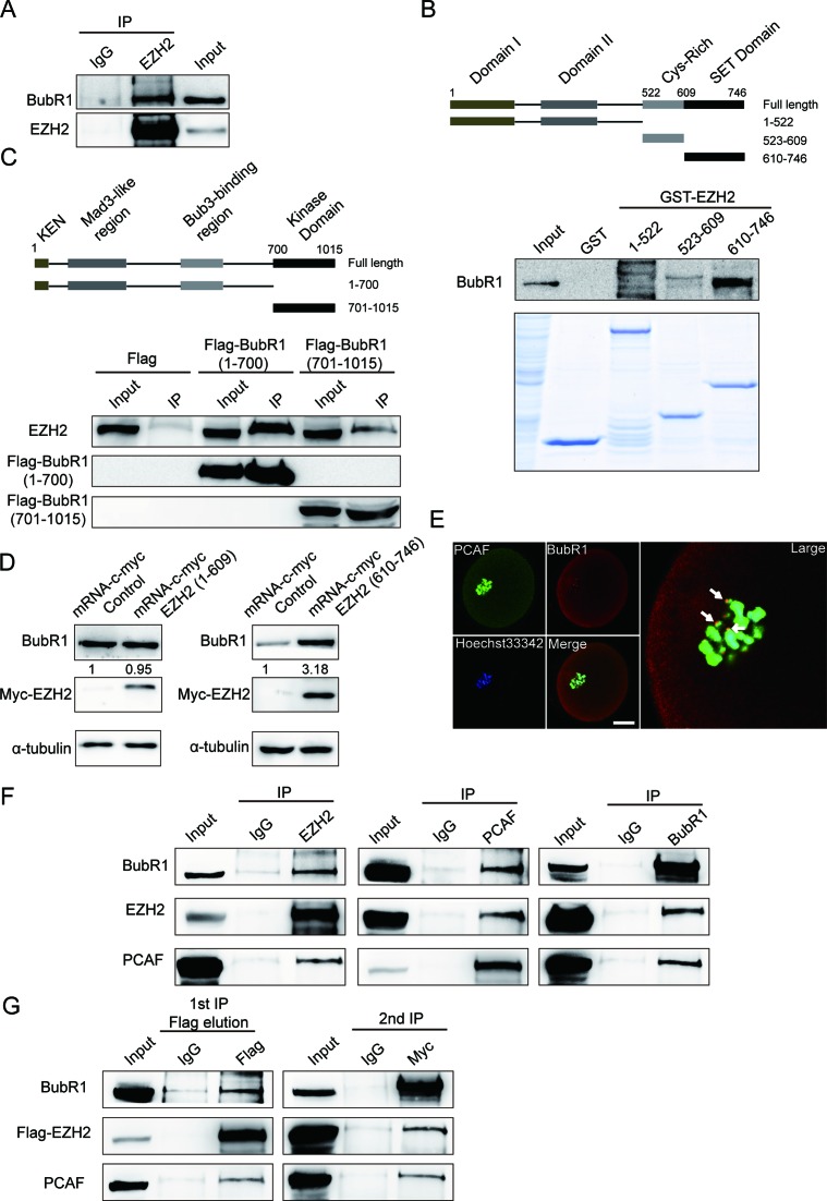
Figure 6.
EZH2 stabilizes BubR1 by directly interacting with BubR1 and also forms a complex with BubR1 and PCAF in mouse oocytes. (A) EZH2 interacts with BubR1 in vivo. BubR1 was co-immunoprecipitated from extracts of approximately 2000 MII oocytes with an EZH2 antibody (lane 2) or with control IgG (lane 1) and 50 MII oocytes were used as input (lane 3). (B) EZH2 SET domain interacts with BubR1. Upper panel: domain structure of EZH2. EZH2 domains with aa 1–522, 523–609 and 610–746 were fused with GST and were expressed in Escherichia coli. Lower panel: an in vitro GST pull-down assay was performed using the above purified GST-EZH2 fragments, and NIH3T3 cell lysates were used as source of BubR1 protein. Protein interaction was detected by western blot analysis with a BubR1 antibody. (C) The N-terminus of BubR1 interacts with EZH2. Upper panel: domain structure of BubR1. Lower panel: NIH3T3 cells were transfected with Flag-BubR1 N-terminus (1-700) and Flag-BubR1 kinase domain (701-1052) vectors and then co-IP assays were performed with Flag-M2 beads. Protein interaction was detected by western blot analysis with an EZH2 antibody. Result showed that the N-terminus of BubR1 interacts with EZH2. (D) BubR1 protein level is raised by the SET domain of EZH2. Left panel: BubR1 protein level could not be raised by EZH2 N-terminal 1–609. C-myc-EZH2:1-609 mRNA was in vitro transcribed and then microinjected into the oocytes for 3 h, followed by transferring to M2 media for 14 h. Experiment was controlled by microinjection of unrelated mRNA. The expression of c-myc-EZH2: 1–609 was detected by a Myc antibody and BubR1 was examined by a BubR1 antibody. Right panel: BubR1 protein level is raised by EZH2 C-terminal 610–746. C-myc-EZH2:610-746 mRNA was in vitro transcribed and then microinjected into the oocytes for 3 h, controlled by microinjection of unrelated mRNA. The expression of c-myc-EZH2:610-746 was detected by a Myc antibody and BubR1 was examined by a BubR1 antibody. (E) PCAF colocalizes with BubR1 at kinetochores in an oocyte. Oocytes were stained with a PCAF antibody (green), BubR1 was stained with a goat pab (red) and chromosome DNAs were stained by Hoechst 33342. Arrowheads indicated examples of colocalization of PCAF and BubR1 at kinetochores. Scale bar = 20 μm. (F) EZH2, BubR1 and PCAF form a complex in mouse oocytes. A panel of co-IP assays using oocyte lysates were performed using EZH2, PCAF or BubR1 antibodies separately and were detected using indicated antibodies. (G) EZH2 forms a complex with BubR1 and PCAF in mammalian cells. Total lysates were extracted from NIH3T3 cells co-transfected with Flag-EZH2 and Myc-BubR1 expression vectors, then sequential co-IPs were performed. The first co-IP was performed using FLAG-M2 beads or mouse immunoglobin IgG to immunoprecipitate EZH2. The eluate from the first co-IP was subjected for the second co-IP with an anti-Myc antibody or rabbit IgG to immunoprecipitate BubR1. The detection of immunoprecipitates from the sequential co-IPs was examined by Western blot analysis using indicated antibodies.

