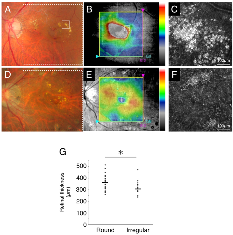
Figure 2. Retinal thickness in regions associated with the two types of hard exudates.
Case 27 with branch retinal vein occlusion (round type, A–C) and case 3 with diabetic retinopathy (irregular type, D–F). (A,D) indicate the color fundus photograph (white solid squares: imaged region of adaptive optics scanning laser ophthalmoscopy (AO-SLO), white dotted squares: imaged region of spectral domain optical coherence tomography (SD-OCT) in B and E, respectively). (B,E) indicate SD-OCT image with the macular cube protocol (128 horizontal line raster with 512 A-scan each, within a 6 × 6 mm2 area). (B,E) Retinal thickness (from ILM to RPE) was displayed geographically as a false-color topographic map. White solid squares in B and E indicate imaged region of AO-SLO in C and F, respectively. AO-SLO image shows accumulation of spherical particles (C) and irregular shaped foci (F). (G) Retinal thickness in the region associated with round type of hard exudates is significantly thicker than that with irregular type of hard exudates (n = 42, P = 0.02, the Wilcoxon matched-pairs signed-ranks tests).

