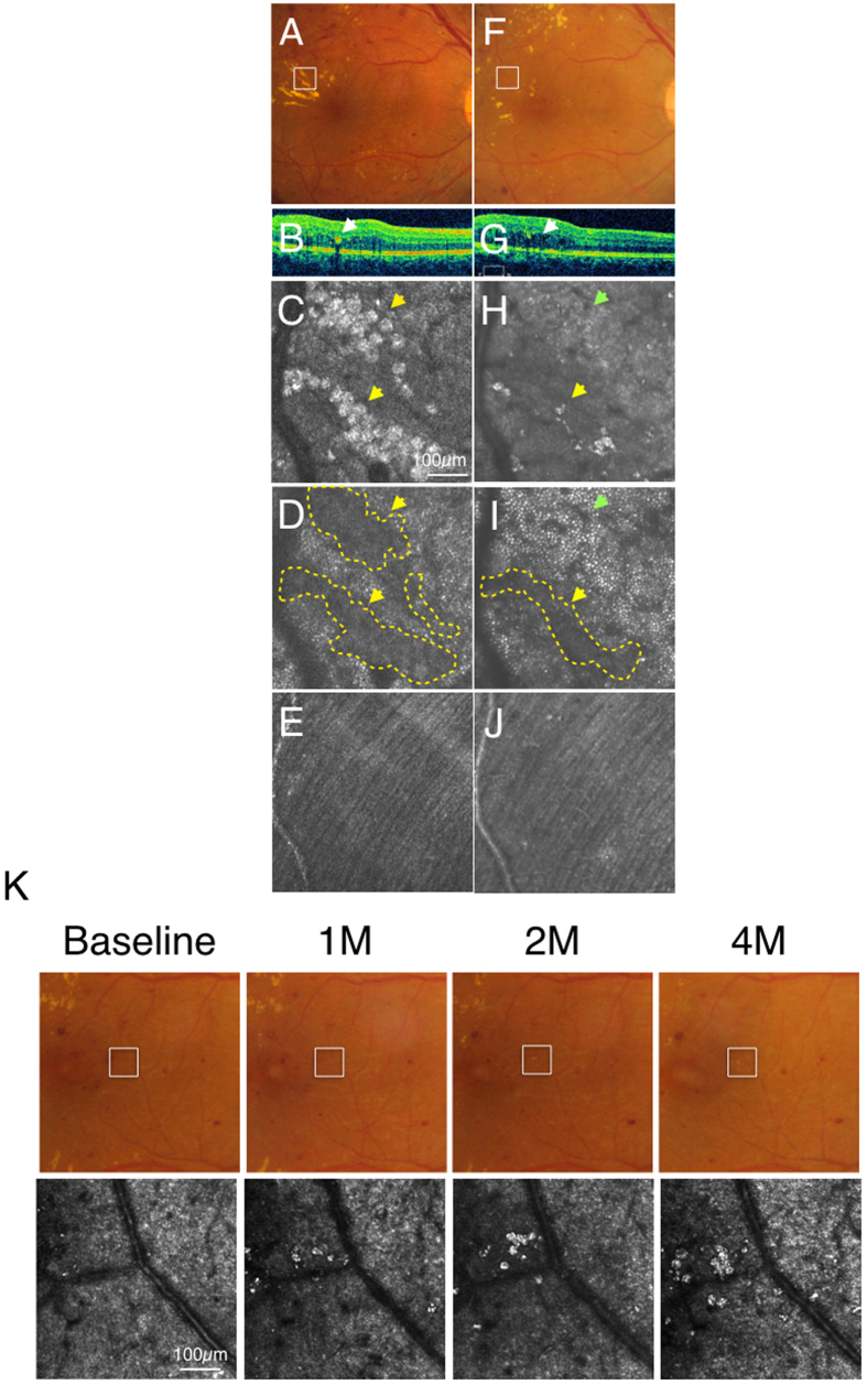
Figure 5.
Case 1 (diabetic retinopathy): Color fundus photograph (A) and spectral domain optical coherence tomography (SD-OCT) (B) showed hard exudates in the outer plexiform layer and the outer nuclear layer (arrows). Adaptive optics scanning laser ophthalmoscopy (AO-SLO) focused and imaged layer with hard exudates (C), photoreceptor layer (D) and nerve fiber layer (E). When focusing at the layer with hard exudates, hard exudates could be observed as hyper-reflective objects (yellow arrows) at the intermediate layer (C). When focused at the photoreceptor layer, dark regions (yellow arrows, yellow dotted areas) could be seen corresponding to the shadow of hard exudates (D). Nerve fiber layer was not affected by hard exudates (E). After one year and four months, color fundus photograph (F), SD-OCT (G) and AO-SLO (H) showed withdrawal of hard exudates. Some dark spots could be still observed at the photoreceptor level (yellow arrow, yellow dotted area) and a cone mosaic could be observed in an area where a dark spot was observed at the first examination (green arrow) (I). Nerve fiber layer is still intact (J). (K) Formation of hard exudates in different regions. AO-SLO image and color fundus photograph with diabetic retinopathy at the first examination, one month later, two months later and four months later. White squares in color fundus photograph indicate imaged regions in AO-SLO. The number of spherical particles increased gradually and accumulated over time. In the color photographs, the spherical particles could be detected as hard exudates.

