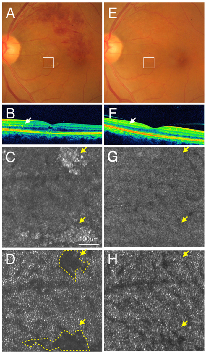
Figure 6.
Case 24 (branch retinal vein occlusion): color fundus photograph (A) and SD-OCT (B) showed hard exudates in the outer plexiform layer (Arrows). Adaptive optics scanning laser ophthalmoscopy (AO-SLO) focused and imaged layer with hard exudates (C) and photoreceptor layer (D). When focused at the layer with hard exudates, hard exudates could be observed as hyper-reflective objects (yellow arrows) (C). When focused at the photoreceptor layer, dark areas (yellow arrows, yellow dotted areas) could be observed corresponding to the shadow of hard exudates (D). After one year and one month, color fundus photograph (E), spectral domain optical coherence tomography (SD-OCT) (F) and AO-SLO (G) showed withdrawal of hard exudates. A cone mosaic could be observed in an area where a dark spot was observed at the first examination (H). Yellow arrows indicate areas where hard exudates could be observed at the first examinations (G,H).

