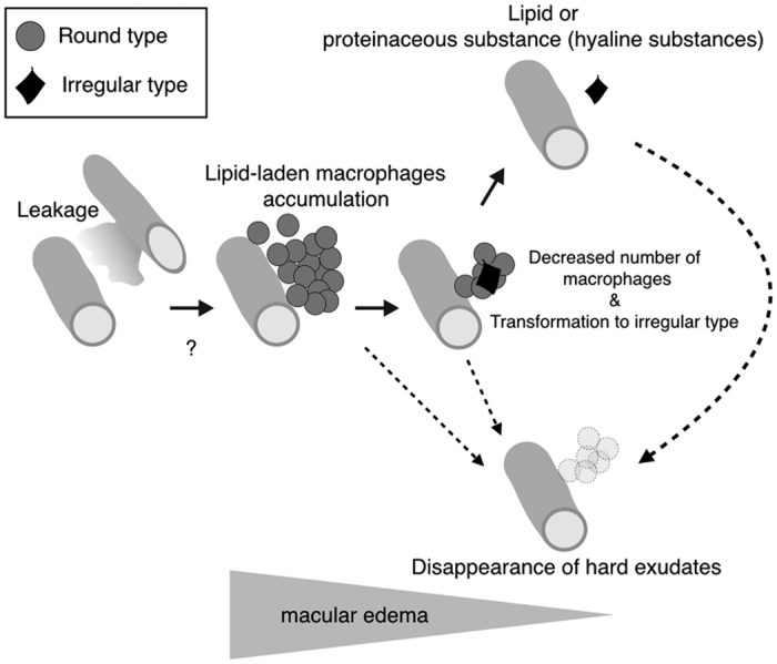
Figure 7. A hypothesized possible model of macrophage-related hard exudates clearance.

High resolution imaging using adaptive optics scanning laser ophthalmoscopy (AO-SLO) shows two types of hard exudates. One consists of macrophages (round type) and the other could be lipid or proteinaceous materials (hyaline substances) (irregular type), respectively, as shown in Fig. 1. In retinal vascular diseases, lipid or proteinaceous materials leak from retinal vessels resulting in macular edema (Fig. 2). During the process of macular edema formation, macrophages accumulate and phagocytose leaked lipid or proteinaceous materials as shown in Fig. 5. This step could be the formation of hard exudates. Upon completion of lipid phagocytosis by macrophages, the number of macrophages then decreases (Fig. 4). If this phagocytosis is accomplished in a physiological manner, hard exudates as well as macular edema could disappear (Figs 4 and 5). If this step was dysfunctional, free lipid or protein derived from lysed phagocytes (the irregular type) could remain. However, it is unknown if irregular hard exudates can disappear without macrophages. Furthermore, it is unclear if the round hard exudates could disappear without transformation to the irregular type.
