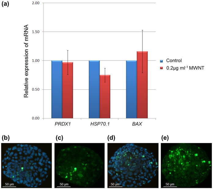
Figure 2. Representative images of TUNEL-labeled nuclei and the relative expression of genes in bovine blastocysts cultured with MWNTs (0.2 μg ml−1) for 72 h.
(a) Shows the expression of PRDX1, HSP70.1 and BAX genes in bovine blastocysts cultured without MWNTs (control group) and with MWNTs (0.2μg ml-1). MWNTs (0.2μg ml-1) compared with the control group (relative expression=1.00) (p>0.05; mean±S.E.M.). (b,c) Show the total number of cells (blue fluorescence) and the number of apoptotic cells (green fluorescence), respectively, from blastocysts of the control group; (d,e) Show the total cell number and apoptotic cells, respectively, from blastocysts of cells cultured with MWNTs (optical microscopy fluorescence with 100x magnification).

