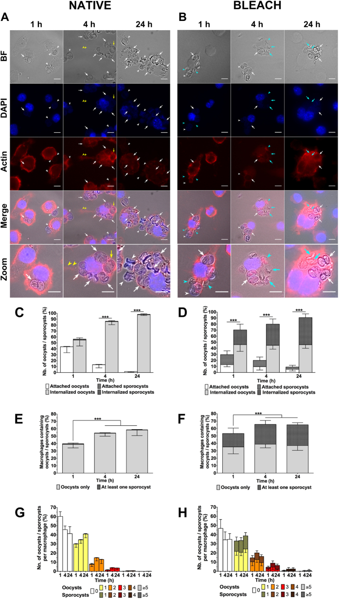
Figure 2. Internalisation of native or bleach-treated oocysts T. gondii oocysts by RAW macrophages.
(A,B) Macrophage cells were incubated with native or bleach-treated oocysts at a ratio of 1:1 for 1, 4 and 24 h at 37 °C and then fixed and stained for macrophage nucleus (blue) and actin cytoskeleton (red) identification. Actin staining allowed to distinguish internalised (white arrow) from attached (white arrowhead) oocysts, in particular freshly internalised ones (yellow arrow), and additionally long pseudopod-like extensions of the macrophage cytoplasm (yellow arrowhead). Light blue arrowheads and arrows denoted sporocysts that adhered to or were internalised by macrophages, respectively. Scale bars: 10 μm. Internalisation assays allowed to calculate (C,D) the number of oocysts/sporocysts attached to or internalised by macrophages, (E,F) the number of macrophages containing oocysts/sporocysts, and (G,H) the number of internalised oocysts/sporocysts per macrophage. ***p < 0.001.

