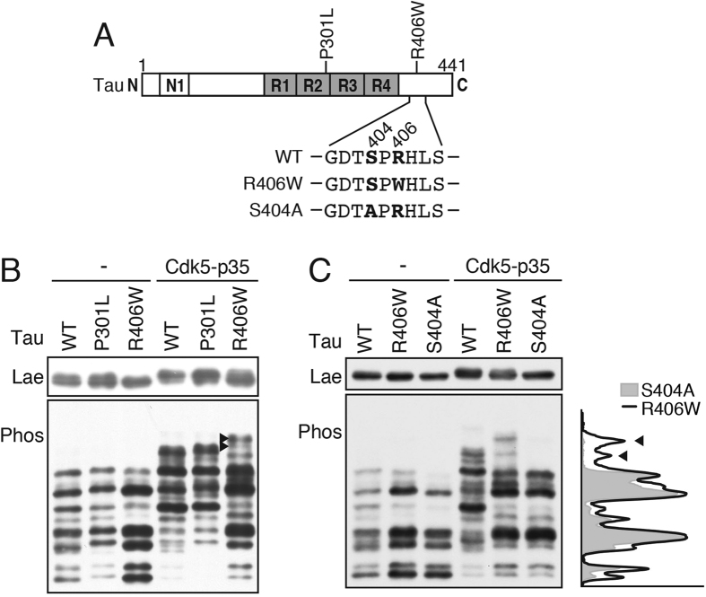
Figure 5. Phosphorylation of tau P301L and R406W mutants by Cdk5-p35.
(A) Positions of P301L and R406W mutations in the tau molecule and amino acid sequences 401–410 of tau WT and R406W and S404A. The Ser404 phosphorylation site and Arg406 mutation site are shown with bold letters. (B) Phos-tag SDS-PAGE analysis of tau WT, P301L and R406W expressed in COS-7 cells with or without co-expression of Cdk5-p35. (C) Comparison of banding pattern of R406W and S404A expressed in COS-7 cells with or without Cdk5-p35. Tau was detected by immunoblotting with Tau5. The upper is Laemmli’s SDS-PAGE and the lower is Phos-tag SDS-PAGE. Densitometric scans of R406W (black line) and S404A (light gray) in the presence of Cdk5-p35 are shown in the right side of the blot. Arrows indicate the bands specifically detected with R406W. Immunoblottings of tau after Laemmli’s SDS-PAGE were performed under the same experimental conditions as an example of the uncropped image, which is provided in Supplemental Fig. 3a.

