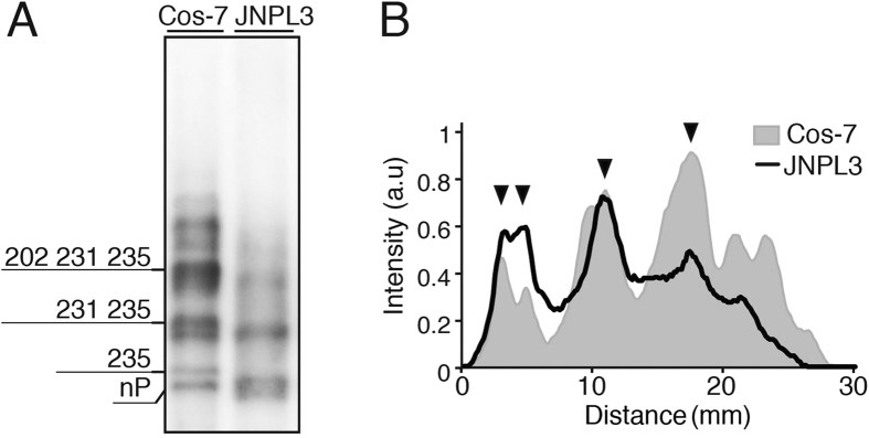
Figure 7. Comparison of phosphorylation profiles of tau expressed in mouse brain and COS-7 cells by Phos-tag SDS-PAGE.
(A) Immunoblotting of human tau WT (0N4R) expressed in COS-7 cells (left lane) and tau P301L (0N4R) in JNPL3 transgenic mouse (right lane) with Tau5 after Phos-tag SDS-PAGE. Phosphorylation sites of major tau bands expressed in COS-7 cells are indicated. (B) Densitometric comparison of the banding patterns between tau in COS-7 cells and mouse brain.

