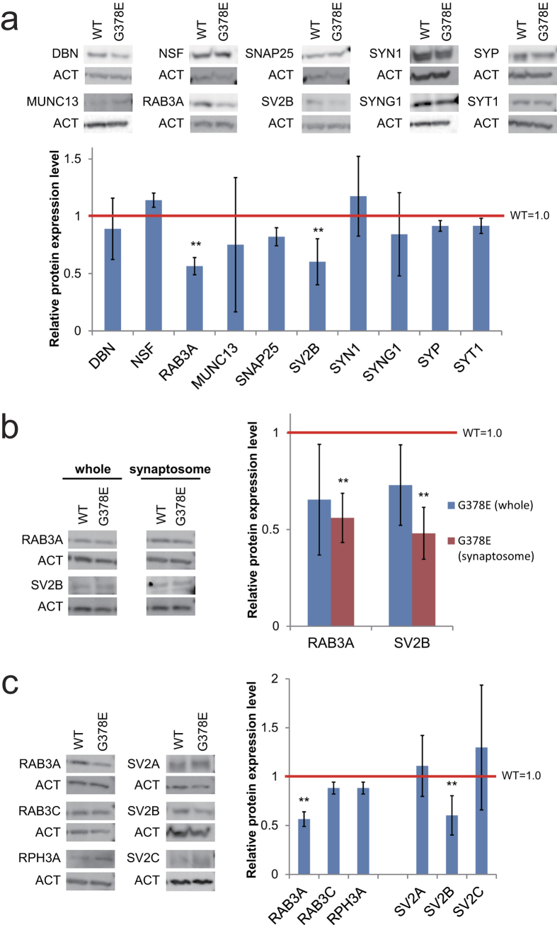
Figure 1. The decrease of RAB3A and SV2B protein levels in the pre-synapses of PS1-G378E neurons.
Immunoblot analyses of the pre-synaptic proteins in the synaptosomes of PS1-G378E neurons (a) RAB3A and SV2B proteins in the whole fraction or synaptosomes of PS1-G378E neurons (b) and the RAB3 and SV2 family proteins in synaptosomes (c). β-actin (ACT) was used as an internal control. Each protein level in the PS1-WT neurons was defined as 1.0. **P < 0.01, as determined by Mann–Whitney U test. Neural differentiation and subsequent synaptosome preparation were independently performed four times (n = 4). Supplementary Fig. S4 shows that isolation of synaptosomes was successfully carried out in all preparations. Mean ± SD. WT, PS1-wild type neurons; G378E, PS1-G378E neurons; DBN, drebrin; NSF, N-ethylmaleimide sensitive factor; RPH3A, rabphilin 3A; SNAP25, synaptosome associated protein 25 kDa; SV2, synaptic vesicle glycoprotein 2; SYN1, synapsin 1; SYNG1, synaptogyrin 1; SYT1, synaptotagmin 1; SYP, synaptophysin.

