Fig. 2.

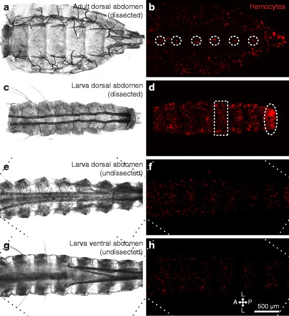
Sessile hemocyte distributions in the abdomen differ between larvae and adults. a, b Dissected adult dorsal abdomen under bright-field (a) and fluorescence illumination (b). Hemocytes (CM-DiI; red) form paired aggregates at the periostial regions of the heart (circles). c, d Dissected fourth instar larval dorsal abdomen under bright-field (c) and fluorescence illumination (d). Hemocytes form segmental bands in each abdominal segment (e.g., rectangle) and are also concentrated in the eighth abdominal segment (oval). e–h Undissected larval abdomen imaged from the dorsal (e, f) and ventral (g, h) views using bright-field (e, g) and fluorescence illumination (f, h). Hemocytes show a segmental banding pattern that encircles the abdomen in each abdominal segment. Diagonal lines in panels e–h denote the edges of rotated images. Directional arrows: A anterior, P posterior, L lateral
