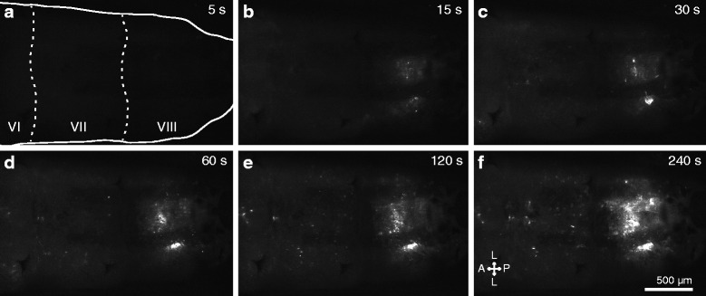
Fig. 5.

E. coli injected into the larval hemocoel rapidly aggregate in the tracheal tufts. a–f Still images from an intravital video (Additional file 4: Movie S2) recorded via fluorescence imaging through the posterior portion of the dorsal cuticle of a larva. GFP-E. coli (white) begin to accumulate at the tracheal tufts within 15 s after treatment. The still images shown were extracted at 5, 15, 30, 60, 120, and 240 s post-treatment. The first panel outlines the larva and marks abdominal segments 6–8 (VI–VIII). Directional arrows: A anterior, P posterior, L lateral
