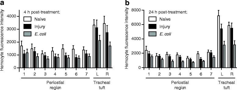
Fig. 9.

Hemocytes are more abundant in the tracheal tufts, but do not increase in response to E. coli infection. a Mean intensity of CM-DiI-stained hemocytes in the periostial regions (1–7) and the left (L) and right (R) tracheal tufts of naïve, injured, and GFP-E. coli-treated larvae at 4 h post-treatment. Hemocyte intensity was not significantly affected by treatment (RM 2 W ANOVA P = 0.1123) but was significantly higher in the tracheal tufts compared to the periostial regions in all three groups (Šidák’s P ≤ 0.0312 for 39 out of 42 comparisons). b Mean intensity of CM-DiI stained hemocytes in the periostial regions and tracheal tufts of naïve, injured, and GFP-E. coli-treated larvae at 24 h post-treatment. Hemocyte intensity was significantly affected by treatment (RM 2 W ANOVA P = 0.0012), with hemocyte intensities from GFP-E. coli-treated larvae being significantly lower than those of both naïve and injured larvae (Šidák’s P < 0.0001 for all comparisons). Hemocyte intensity was significantly higher in the tracheal tufts compared to the periostial regions in all three groups (Šidák’s P ≤ 0.0003 for all comparisons). Whiskers denote the SEM. For a graphical presentation of how the ROIs were constructed, see Additional file 7: Figure S5C
