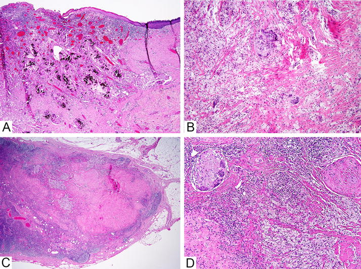
Figure 2. Lymph node with complete pathological response to neoadjuvant chemotherapy.

This patient had complete pathological response to neoadjuvant chemotherapy. A. Low power image of primary tumour bed in the breast. B. Medium power image of primary tumour bed in the breast. C. The lymph node shows marked fibrosis at low power. D. Foamy macrophages and multinucleated giant cells are present in the lymph node. The features are morphologically similar to those seen in the tumour bed in the breast.
