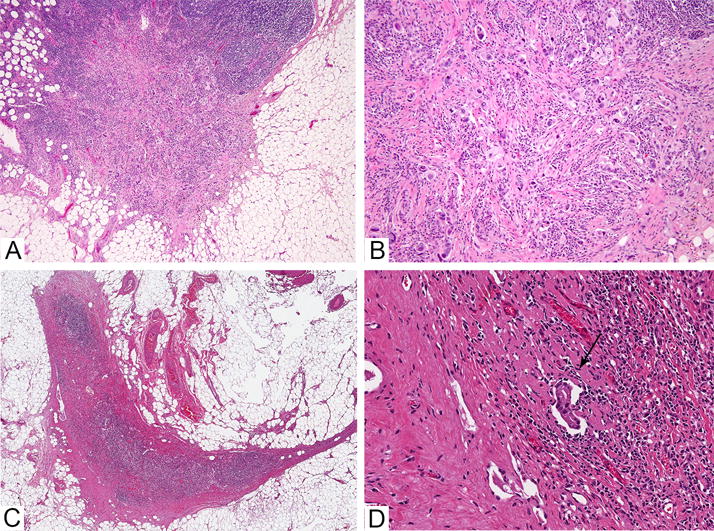
Figure 3. Lymph nodes with residual metastatic carcinoma post neoadjuvant chemotherapy.

A. Low power image of lymph node with residual metastastic carcinoma composed of single cells in desmoplastic stroma. Extracapsular extension is present. B. Higher power image of the same lymph node. C. Low power image of a lymph node with a subtle focus of residual carcinoma after neoadjuvant chemotherapy, seen at higher power in image D (see arrow).
