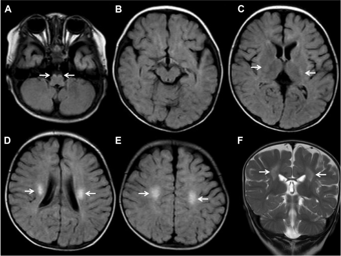
Figure 1.
Axial FLAIR (A–E) and coronal T2-weighted (F) MRI images.
Notes: Figure shows high intensities in the bilateral corticospinal tracts from the centrum semiovale through the posterior internal capsule to the brainstem (white arrows). There are no abnormalities of the dentate and cerebellar white matter.
Abbreviations: FLAIR, fluid-attenuated inversion recovery; MRI, magnetic resonance imaging.

