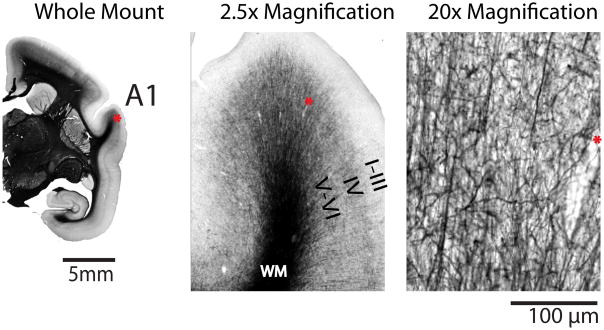
Figure 1.
Representative cortical myelination in the marmoset. A 40 μm-thick coronal histological section stained for myelin using a modified Gallyas silver staining method. At the whole mount level of magnification showing half of the brain, distinct dense areas of staining are seen which correspond to specific cortical areas (for example, the primary auditory cortex (A1)). At 2.5 times magnification, dark stained fibres can been seen running vertically from the white matter (WM) through Layers VI and V. At 20 times magnification in Layer IV, the fibres are arborized and vertical and horizontal stained branches can be seen. There is little myelin staining through Layers III -I. The same pattern is seen in other myelinated areas of the marmoset cortex, save for the primary visual cortex (V1) where the density of myelination is highest in Layer IV (the Stripe of Gennari). The asterix denotes the same blood vessel at each magnification.

