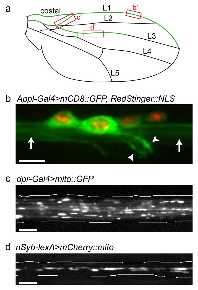
Figure 1. Imaging organelle transport in the nervous system of the adult Drosophila wing.
(a) Cartoon of the Drosophila wing showing the position of the costal and longitudinal veins (L1–L5). The nervous system is highlighted in green. Boxes highlight regions imaged in b–d. (b) Cell bodies and processes of L1 vein neurons. Green, cell membranes; red, nuclei (genotype is Appl-Gal4>mCD8::GFP, RedStinger::NLS). Arrowheads, dendrites; arrows, bundled axons. (c) Still image of GFP-labelled mitochondria in axons of the wing arch region of a dpr-Gal4>mito::GFP fly one day after eclosion (from Supplementary Video 2). (d) Still image of mCherry-labelled mitochondria in axons of the L3 vein of a nSyb-lexA>mCherry::mito fly two days after eclosion (from Supplementary Video 3). Images in b–d were captured with spinning disk microscopy using the procedures described in the protocol (b, z-stack projection; c and d, single focal planes). Scale bars: 5 μm.

