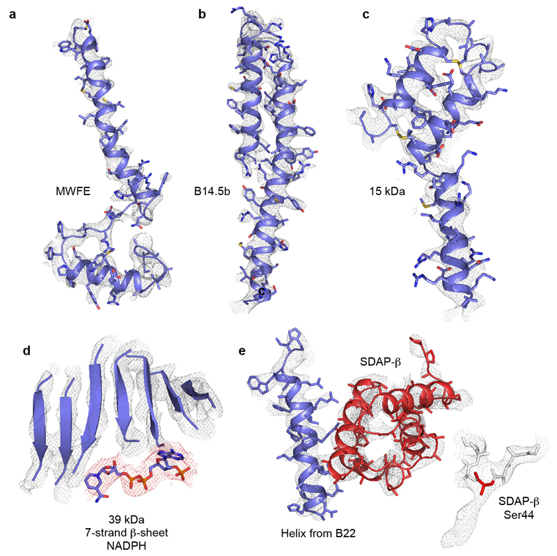
ED Figure 4. Example regions of the cryoEM density map for the supernumerary subunits, and the model fitted to the map.
a) Subunit MWFE, containing one TMH. b) Subunit B14.5, containing two TMHs. The N- and C-terminal loops are not shown. c) 15 kDa subunit on the IMS face, containing a CHCH domain with two disulphide bonds. The N- and C-terminal loops are not shown. d) The seven-strand β-sheet in the 39 kDa subunit, showing the separation of the strands, and the bound nucleotide (red density) modelled as NADPH. e) Helix 1, one of the arginine-rich helices, in B22, and SDAP-β, on the matrix side of the tip of the membrane domain. Inset: the weak density attached to Ser44 in SDAP-β attributed to the attached pantetheine-4'-phosphate group (side chain of Ser44 not shown).

