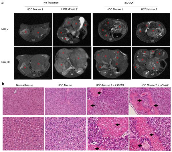
Figure 2.
Representative MRI and H & E staining to show inCVAX-generated tumor damage. (a) MRI scan: Tumor mass in tumor-bearing mouse prior and post inCVAX treatment was monitored by MRI. Stars showed tumor mass. (b) H&E staining: Sections of liver tissues from normal mouse and tumor tissues in large tumor-bearing mice with and without inCVAX were stained. Arrows showed inCVAX-induced obvious tumor necrosis. Upper panel: low magnification; Lower panel: high magnification.

