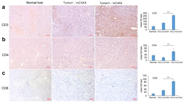
Figure 4.
Effect of inCVAX on T cell infiltration into tumors. Immunohistochemical staining of CD3 (a), CD4 (b), and CD8 (c) was performed in normal liver tissues from control syngeneic wild type C57BL/6 mice and tumor tissues from large HCC-bearing mice with and without inCVAX. Few CD3+, CD4+, and CD8+ T cells were observed in normal mice and large HCC-bearing mice with equivalent level of CD4 and slight increase in HCC-bearing mice. In contrast, inCVAX led to high numbers of CD3+, CD4+, and CD8+ T cells detected in tumor tissues. The average liver- or tumor-infiltrated T cells per 100 fields were calculated. 100× magnification, n=3, *p<0.05, ** p<0.01, error bars represent mean ± SDs.

