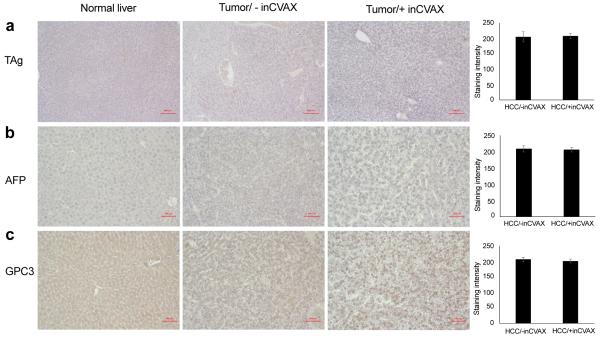
Figure 6.
Influence of inCVAX on tumor antigens. Immunohistochemical staining of TAg (a), GPC-3 (b), and α-AFP (c) were performed in normal liver tissues from control syngeneic wild type C57BL/6 mice and tumor tissues from large HCC-bearing mice with and without inCVAX. In contrast to normal mice, specific expression of TAg and obviously increased production of GPC-3 and α-AFP were detected in tumor-bearing mice in photomicrographs at 100x magnification. There were no significant expression changes of three antigens detected in HCC-bearing mice after inCVAX treatment.

