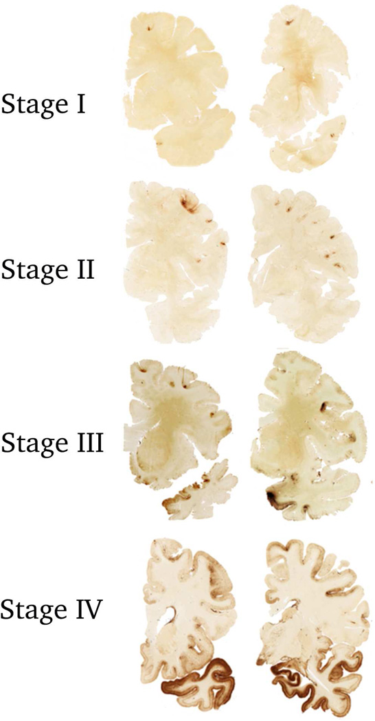
Figure 1. Stages of Hyperphosphorylated Tau Pathology in CTE.
In stage I CTE, p-tau pathology is restricted to isolated foci in the cerebral cortex, the focal lesions consist of perivascular accumulation of p-tau as neuronal and astrocytic inclusions, with NFTs and dot-like structures.
In stage II CTE, there are multiple p-tau lesions typically found at the depths of the cerebral sulci In stage III CTE, p-tau pathology is widespread in the cortex and the amygdala, hippocampus and entorhinal cortex show neurofibrillary pathology.
In stage IV CTE, there is widespread severe p-tau pathology affecting most regions of the cerebral cortex and the medial temporal lobe, with sparing of the calcarine crtex. All images, CP-13 immunostained 50 μ tissue sections.

