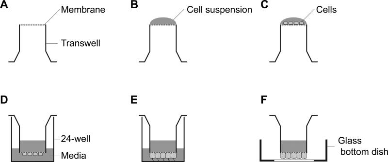
Figure 1. Sample preparation for imaging IFT in mammalian primary cilia.
(A) The transwell cup is placed upside down in a humid sterile chamber. (B) Put 50 μL of the cell suspension (1 × 10⁴ cells) on the membrane of the inverted transwell cup. (C) Culture cells for 3 hours at 37°C in 5% CO2. (D) The transwell cup is transferred to 24-well plate and media add into the transwell cup and the 24-well. (E) Culture cells until 90% confluency. (F) Put the transwell cup on a 35-mm glass bottom dish for imaging. We call it transwell imaging chamber.

