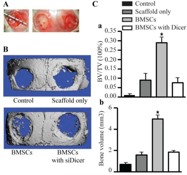
Figure 6.
Bone regeneration of calvarial bone critical-size defects (CSDs) in mice. (A) The non-healing full thickness defect of 4 mm in diameter was made in both sides of the cranial bone and filled with a silk scaffold seeded with gene-modified BMSCs. (B) Representative pictures of the morphology of the newly formed bone was reconstructed using micro-CT imaging in 4 groups (control, defects unfilled; scaffolds only, defects filled with scaffolds alone; BMSCs, defects filled with scaffolds seeded with BMSCs; BMSCs with siDicer, defects filled with scaffolds seeded with BMSCs transfected with siRNA targeting Dicer). (C) To quantify the new bone regeneration within the calvarial defects, the ratio of bone volume to total volume in region of interest (BV/TV) (a), bone volume (b) were measured in microCT scanned images 4 weeks after surgery. These data are expressed as the mean ± SD (n=4). * p < 0.05, versus control.

