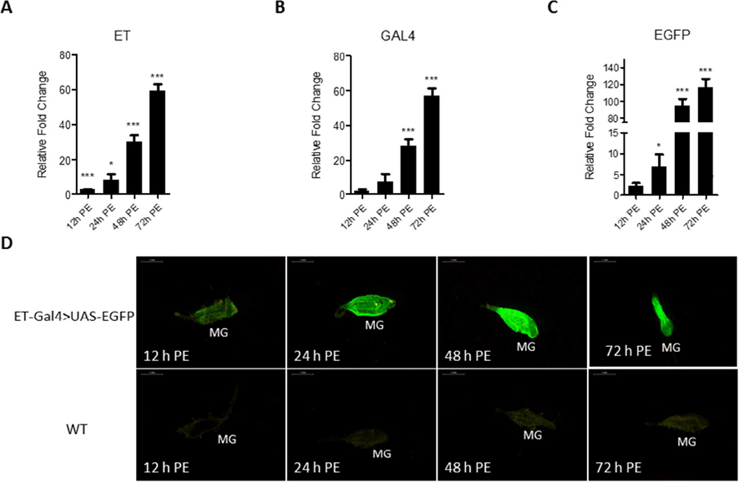
Figure 3.
Expression of the ET-Gal4>UAS-EGFP transgene in the Ae. aegypti hybrid line. (A–C) The time course of transcript abundance in midguts of the ET-Gal4>UAS-EGFP hybrid transgenic female mosquitoes. RNA samples were extracted from isolated midguts of female mosquitoes at different time points: 12 h PE, 24 h PE, 48 h PE, and 72 h PE. Expression patterns of Gal4 and EGFP were highly similar to that of the endogenous ET. Relative fold changes were established by comparing transcript levels of each sample with 0 h PE mosquito sample. The abundance of 0 h is represented as 1.0, with corresponding adjustments for other time points. Values represent average ± s.e.m. from three combined biological replicates. *p < 0.05; **p < 0.01; ***p < 0.001 (t-test). (D) Images of midguts (MG) of the ET-Gal4>UAS-EGFP transgenic hybrid female mosquitoes with EGFP signal at several time points during the PE phase. Samples from wild type (WT) mosquitoes served as negative control. Images were obtained using a Leica M165FC fluorescent stereomicroscope with LAS V4.0 software. Scale bar: 1 mm.

