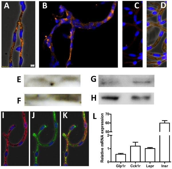
Fig. 1. CCK-1R is expressed in rat brain microvessels.
CCK-1R immunoreactivity was detected in freshly-isolated brain microvessels via fluorescence microscopy (A-B, orange/Alexa Fluor® 594, 40X magnification), imaged with phase contrast (A) and without phase contrast (B). Nuclei were stained with DAPI (blue). No immunoreactivity was observed in control IHC experiments that omitted primary antibody (C-D); images were taken with phase contrast (C) and without phase contrast (D), for comparison. Western blotting was performed with protein lysates from brain microvessel isolates (E) and choroid plexuses (F) using the Santa Cruz CCK- 1R antibody (47-50 kDa) and using the LS Biosciences (~90 kDa) antibody in separate blots of brain microvessel isolates (G) and choroid plexuses (H). Insulin receptor protein expression was detected in brain microvessels (I) and vessels were co-immunoblotted with a rat brain microvessel antibody (J, alone and K, together). CCK-1R mRNA expression (Cck1r) was also detected in rat brain microvessel isolates (L) at levels comparable to that of the full leptin receptor transcript (Lepr). Other genes examined included the GLP-1 receptor (Glp1r) and insulin receptor (Insr). n=6, ± SEM.

