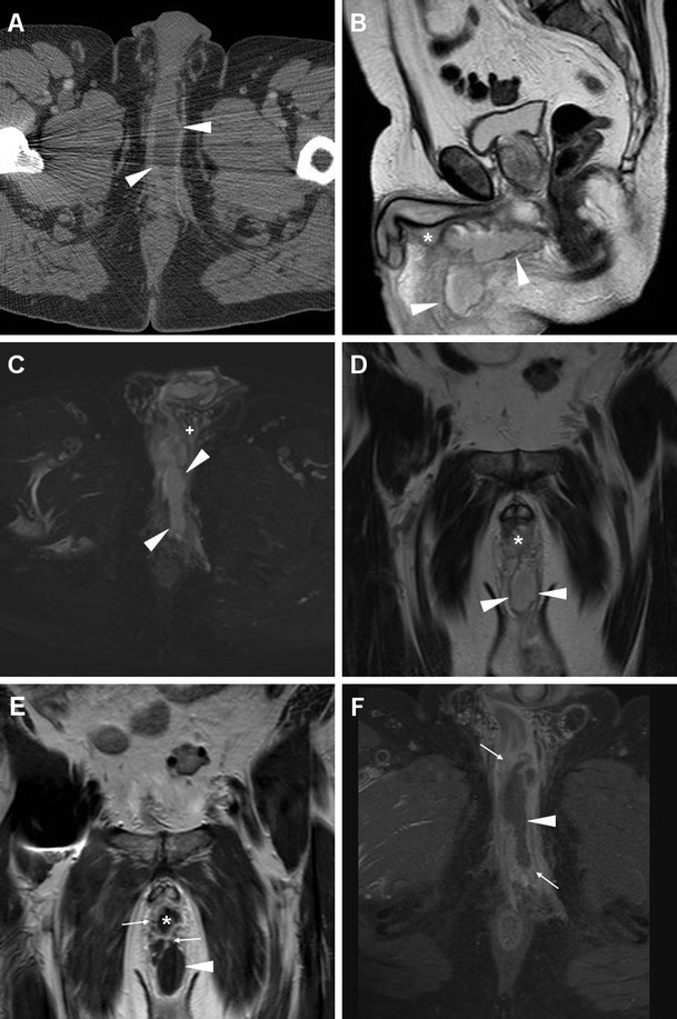
Fig. 15.

Urethral infection complicated by penile and perineal abscess in a 53-year-old man with tender, inflamed perineal swelling despite antibiotics. Infection was initially detected at contrast-enhanced CT (a) as an elongated midline abscess with peripheral enhancement (arrowheads) and internal fluid. MRI showed corresponding inhomogeneous fluid-like content on T2-weighted sequences (b–d), with surrounding inflammatory stranding (+) and strong contrast enhancement in the abscess walls (arrowheads in e, f). The infected corpus spongiosum (*) showed similar signal features. Surgical evacuation was required to relieve the abscess
