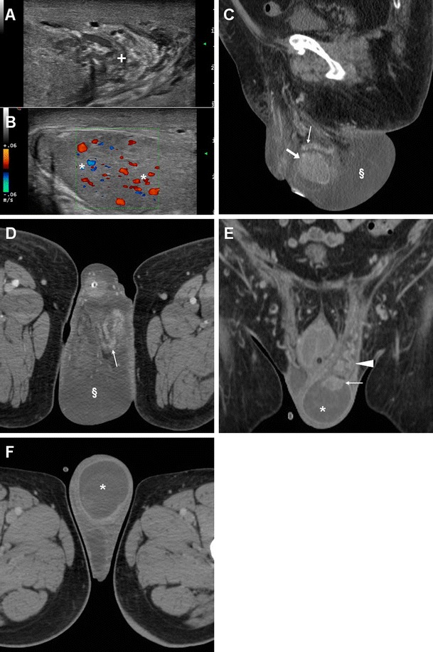
Fig. 18.

Surgically confirmed epididymo-orchitis with pyocele in a 72-year-old diabetic man with haematuria and enlarged left scrotum, history of transurethral resection of bladder carcinoma and bladder neck stricture treated by long-term catheterisation. Ultrasound revealed ipsilateral enlarged inhomogeneous epididymal head (+ in a) and hypervascularised testis (* in b). After unsuccessful antibiotic therapy, contrast-enhanced CT (c, d) showed hyperaemic left epididymis (thin arrows) and testis (arrows) compared to contralateral structures, and development of posterior scrotal collection (§). Another surgically proven case of testicular abscess and necrosis in a 59-year-old man with epididymo-orchitis unresponsive to medical therapy: post-contrast CT (e, f) revealed vascular engorgement along the left spermatic cord (arrowhead), faintly enhanced epididymal head (thin arrow in e), and ipsilateral scrotum occupied by fluid-like collection (*) with thin peripheral enhancing rim
