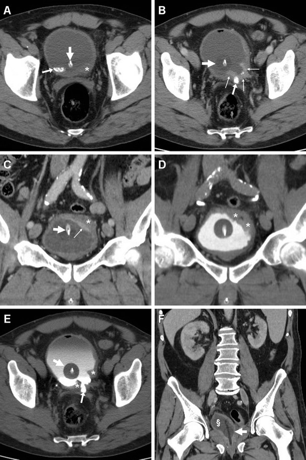
Fig. 7.

Muscle-invasive bladder carcinoma in a 54-year-old man with urolithiasis (arrows) and long-term bladder catheterisation (thick arrows). Unenhanced (a), portal (b, c) and excretory (d, e) phase CT images showed focal solid mural thickening (*) at the left posterolateral bladder wall, with an irregular configuration and positive contrast enhancement (thin arrows). Postoperative status after radical cystectomy (f) with orthotopic neobladder (§) is shown on follow-up CT (f)
