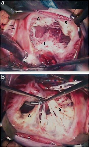
Fig. 1.

Normal surgical anatomy. (a) Superior view of the tricuspid valve shows the anterior leaflet (A), which is the largest; the septal leaflet (S), which is the smallest; and the posterior/inferior leaflet (P). (b) Another view of the tricuspid valve showing the papillary muscles, which are more numerous, smaller and more widely separated than those on the left side of the heart
