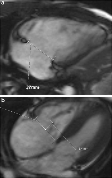
Fig. 11.

Tricuspid regurgitation (TR). (a) Four-chamber steady-state free precession (SSFP) image in a patient with functional TR shows dilation of the annulus > 35 mm. The annulus is also flat and planar. Dilation occurs along the free wall of the tricuspid annulus. The septal wall is intact. (b) Tethering. Four-chamber SSFP MRI image shows distance from the annular plane to coaptation in systole > 8 mm and area > 1.6 cm2, predicting residual TR following surgery
