Fig. 16.

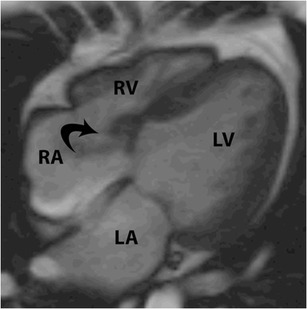
Gerbode defect. Four-chamber steady-state free precession (SSFP) image shows a defect and a shunt extending from the left ventricle (LV) to the right atrium (RA) through a Gerbode defect

Gerbode defect. Four-chamber steady-state free precession (SSFP) image shows a defect and a shunt extending from the left ventricle (LV) to the right atrium (RA) through a Gerbode defect