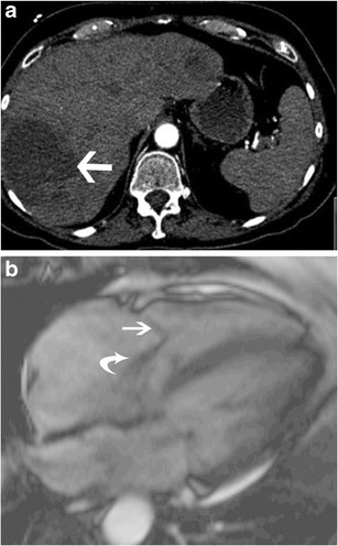
Fig. 20.

Carcinoid. (a) Axial CT of the abdomen shows a metastatic carcinoid tumour (arrow) in the liver. (b) Four-chamber steady-state free precession (SSFP) image shows a thickened tricuspid leaflet (straight arrow) with moderate tricuspid regurgitation (curved arrow). Mitral regurgitation is also visible
