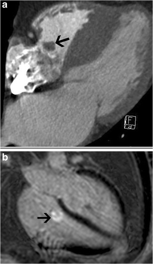
Fig. 23.

Fibroelastoma. (a) Four-chamber CT image shows a small, well-defined mass attached to the septal leaflet of the tricuspid valve (arrow), consistent with a fibroelastoma. (b) Four-chamber delayed-enhancement image in another patient shows heterogeneous enhancement of the tricuspid valve mass, consistent with a fibroelastoma
