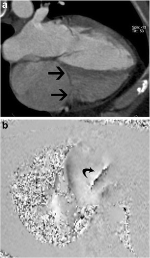
Fig. 6.

Tricuspid stenosis in CT and MRI. (a) Four-chamber reconstructed CT image shows thickening of the tricuspid leaflets (arrows) in a patient with carcinoid and tricuspid stenosis. (b) Four-chamber phase contrast velocity-encoded image shows a high-velocity jet extending across the tricuspid valve, resulting in aliasing (arrow)
