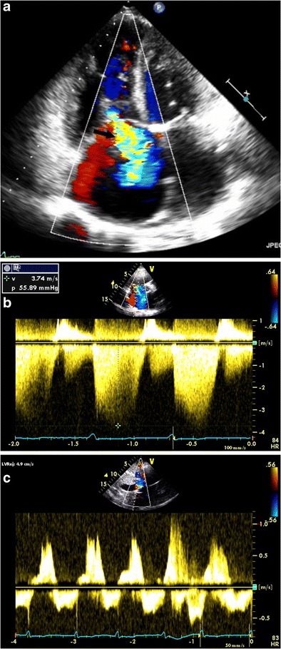
Fig. 7.

Echocardiography of tricuspid regurgitation (TR). (a) Apical four-chamber view with color Doppler demonstrates a markedly enlarged RA and a large TR jet (arrow) coursing along the interatrial septum. (b) Parasternal short-axis view with color Doppler demonstrates elevated RV pressure via peak TR jet velocity. (c) Hepatic vein continuous-wave Doppler with systolic reversal of flow in the setting of severe TR
